Heart attack facts
- A heart attack results when a blood clot completely obstructs a coronary artery supplying blood to the heart muscle and heart muscle dies.
- The blood clot that causes the heart attack usually forms at the site of rupture of an atherosclerotic, cholesterol plaque on the inner wall of a coronary artery.
- The most common symptom of heart attack is chest pain.
- The most common complications of a heart attack are heart failure and ventricular fibrillation.
- The risk factors for atherosclerosis and heart attack include elevatedcholesterol levels, increased blood pressure, tobacco use, diabetes, male gender, and a family history ofheart attacks at an early age.
- Heart attacks are diagnosed with electrocardiograms and measurement of cardiac enzymes in blood.
- Treatment guidelines emphasize treatment at a hospital capable of doing PCI (percutaneous coronary intervention) also termed as stenting or stent placement.
- Early reopening of blocked coronary arteries reduces the amount of damage to the heart and improves the prognosis for a heart attack.
- Medical treatment for heart attacks may include antiplatelet, anticoagulant, and clot dissolving drugs as well as angiotensin converting enzyme (ACE) inhibitors, beta blockers, and oxygen.
- Interventional treatment for heart attacks may include coronary angiography with percutaneous transluminal coronary angioplasty (PTCA), coronary arterystents, and coronary artery bypass grafting (CABG).
- Patients suffering a heart attack are hospitalized for several days to detect heart rhythm disturbances, shortness of breath, and chest pain.
- Further heart attacks can be prevented by aspirin, beta blockers, ACE inhibitors, discontinuing smoking, weight reduction, exercise, good control of blood pressure and diabetes, following a low cholesterol and low saturated fat diet that is high in omega-3-fatty acids, taking multivitamins with an increased amount of folic acid, decreasing LDL cholesterol, and increasing HDL cholesterol.
A heart attack (also known as a myocardial infarction or MI) is the damage and death of heart muscle from the sudden blockage of a coronary artery by a blood clot. Coronary arteries are blood vessels that supply the heart muscle with blood and oxygen. Blockage of a coronary artery deprives the heart muscle of blood and oxygen, causing injury to the heart muscle. Injury to the heart muscle causes chest pain and chest pressure sensation. If blood flow is not restored to the heart muscle within 20 to 40 minutes, irreversible death of the heart muscle will begin to occur. Muscle continues to die for six to eight hours at which time the heart attack usually is "complete." The dead heart muscle is eventually replaced by scar tissue.
What causes a heart attack?
Atherosclerosis
Atherosclerosis is a gradual process by which plaques (collections) of cholesterol are deposited in the walls of arteries. Cholesterol plaques cause hardening of the arterial walls and narrowing of the inner channel (lumen) of the artery. Arteries that are narrowed by atherosclerosis cannot deliver enough blood to maintain normal function of the parts of the body they supply. For example, atherosclerosis of the arteries in the legs causes reduced blood flow to the legs. Reduced blood flow to the legs can lead to pain in the legs whilewalking or exercising, leg ulcers, or a delay in the healing of wounds to the legs. Atherosclerosis of the arteries that furnish blood to the brain can lead to vascular dementia (mental deterioration due to gradual death of brain tissue over many years) or stroke(sudden damage and death of brain tissue).
In many people, atherosclerosis can remain silent (causing no symptoms or health problems) for years or decades. Atherosclerosis can begin as early as the teenage years, but symptoms or health problems usually do not arise until later in adulthood when the arterial narrowing becomes severe. Smoking cigarettes, high blood pressure, elevated cholesterol, anddiabetes mellitus can accelerate atherosclerosis and lead to the earlier onset of symptoms and complications, particularly in those people who have a family history of early atherosclerosis.
Coronary atherosclerosis (or coronary artery disease) refers to the atherosclerosis that causes hardening and narrowing of the coronary arteries. Diseases caused by the reduced blood supply to the heart muscle from coronary atherosclerosis are called coronary heart diseases (CHD). Coronary heart diseases include heart attacks, sudden unexpected death, chest pain (angina), abnormal heart rhythms, and heart failure due to weakening of the heart muscle.
Atherosclerosis and angina pectoris
Angina pectoris (also referred to as angina) is chest pain or pressure that occurs when the blood and oxygen supply to the heart muscle cannot keep up with the needs of the muscle. When coronary arteries are narrowed by more than 50 to 70 percent, the arteries may not be able to increase the supply of blood to the heart muscle during exercise or other periods of high demand for oxygen. An insufficient supply of oxygen to the heart muscle causes angina. Angina that occurs with exercise or exertion is called exertional angina. In some patients, especially in people with diabetes, the progressive decrease in blood flow to the heart may occur without any pain or with just shortness of breath or unusually early fatigue.
Exertional angina usually feels like a pressure, heaviness, squeezing, or aching across the chest. This pain may travel to the neck, jaw, arms, back, or even the teeth, and may be accompanied by shortness of breath, nausea, or a cold sweat. Exertional angina typically lasts from one to 15 minutes and usually is relieved by rest or by placing a tablet ofnitroglycerin under the tongue. Both resting and nitroglycerin decrease the heart muscle's demand for oxygen, thus relieving angina. Exertional angina may be the first warning sign of advanced coronary artery disease. Chest pains that just last a few seconds rarely are due to coronary artery disease.
Angina also can occur at rest. Angina at rest more commonly indicates that a coronary artery has narrowed to such a critical degree that the heart is not receiving enough oxygen even at rest. Angina at rest infrequently may be due to spasm of a coronary artery (a condition called Prinzmetal's or variant angina). Unlike a heart attack, there is no permanent muscle damage with either exertional or rest angina although the angina is a warning sign that there is an increased chance of a heart attack in the future.
Atherosclerosis and heart attack
Occasionally the surface of a cholesterol plaque in a coronary artery may rupture, and a blood clot forms on the surface of the plaque. The clot blocks the flow of blood through the artery and results in a heart attack (see picture below). The cause of rupture that leads to the formation of a clot is largely unknown, but contributing factors may include cigarette smoking or other nicotine exposure, elevated low-density lipoprotein (LDL) cholesterol, elevated levels of blood catecholamines (adrenaline), high blood pressure, and other mechanical and biochemical stimuli.
Unlike exertional or rest angina, heart muscle dies during a heart attack and loss of the muscle is permanent, unless blood flow can be promptly restored, usually within one to six hours.

While heart attacks can occur at any time, more heart attacks occur between 4 A.M. and 10 A.M. because of the higher blood levels of adrenaline released from the adrenal glands during the morning hours. Increased adrenaline, as previously discussed, may contribute to rupture of cholesterol plaques.
Only half of patients who develop heart attacks have warning signs such as exertional angina or rest angina prior to their heart attacks, but these signs may be mild and ignored as unimportant.
What are the symptoms of a heart attack?
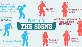
Although chest pain or pressure is the most common symptom of a heart attack, heart attack victims may experience a variety of conditions including:
- Pain, fullness, and/or squeezing sensation of the chest
- Jaw pain, toothache, headache
- Shortness of breath
- Nausea, vomiting, and/or general epigastric (upper middle abdomen) discomfort
- Sweating
- Heartburn and/or indigestion
- Arm pain (more commonly the left arm, but may be either arm)
- Upper back pain
- General malaise (vague feeling of illness)
- No symptoms (Approximately one quarter of all heart attacks are silent, without chest pain or new symptoms. Silent heart attacks are especially common among patients with diabetes mellitus.)
Even though the symptoms of a heart attack at times can be vague and mild, it is important to remember that heart attacks producing no symptoms or only mild symptoms can be just as serious and life-threatening as heart attacks that cause severe chest pain. Too often patients attribute heart attack symptoms to "indigestion," "fatigue," or "stress," and consequently delay seeking prompt medical attention. One cannot overemphasize the importance of seeking prompt medical attention in the presence of new symptoms that suggest a heart attack. Early diagnosis and treatment saves lives, and delays in reaching medical assistance can be fatal. A delay in treatment can lead to permanently reduced function of the heart due to more extensive damage to the heart muscle. Death also may occur as a result of the sudden onset of arrhythmias such as ventricular fibrillation.
What are the complications of a heart attack?
Heart Failure
When a large amount of heart muscle dies, the ability of the heart to pump blood to the rest of the body is diminished, and this can result in heart failure. The body retains fluid, and organs, for example, the kidneys, begin to fail.
Ventricular fibrillation
Injury to heart muscle also can lead to ventricular fibrillation. Ventricular fibrillation occurs when the normal, regular, electrical activation of heart muscle contraction is replaced by chaotic electrical activity that causes the heart to stop beating and pumping blood to the brain and other parts of the body. Permanent brain damage and death can occur unless the flow of blood to the brain is restored within five minutes.
Most of the deaths from heart attacks are caused by ventricular fibrillation of the heart that occurs before the victim of the heart attack can reach an emergency room. Those who reach the emergency room have an excellent prognosis; survival from a heart attack with modern treatment should exceed 90%. The 1% to 10% of heart attack victims who later die frequently had suffered major damage to the heart muscle initially or additional damage at a later time.
Deaths from ventricular fibrillation can be avoided bycardiopulmonary resuscitation (CPR) started within five minutes of the onset of ventricular fibrillation. CPR requiresbreathing for the victim and applying external compression to the chest to squeeze the heart and force it to pump blood. In 2008, the American Heart Association modified the mouth-to-mouth instruction of CPR, and recommends that chest compressions alone are effective if a bystander is reluctant to do mouth-to-mouth. When paramedics arrive, medications and/or an electrical shock (cardioversion) can be administered to convert ventricular fibrillation back to a normal heart rhythm and allow the heart to pump blood normally. Therefore, prompt CPR and a rapid response by paramedics can improve the chances of survival from a heart attack. In addition, many public venues now have automatic external defibrillators (AEDs) that provide the electrical shock needed to restore a normal heart rhythm even before the paramedics arrive. This greatly improves the chances of survival.
What are the risk factors for atherosclerosis and heart attack?
Factors that increase the risk of developing atherosclerosis and heart attacks include increased blood cholesterol, high blood pressure, use of tobacco, diabetes mellitus, male gender (although women may still be very much at risk -- see section at end of article), and a family history of coronary heart disease. While family history and male gender are genetically determined, the other risk factors can be modified through changes in lifestyle and medications.
- High Blood Cholesterol (Hyperlipidemia). A high level of cholesterol in the blood is associated with an increased heart attack risk because cholesterol is the major component of the plaques deposited in arterial walls. Cholesterol, like oil, cannot dissolve in the blood unless it is combined with special proteins called lipoproteins. (Without combining with lipoproteins, cholesterol in the blood would turn into a solid substance.) The cholesterol in blood is either combined with lipoproteins as very low-density lipoproteins (VLDL), low-density lipoproteins (LDL) or high-density lipoproteins (HDL).
The cholesterol that is combined with low-density lipoproteins (LDL cholesterol) is the "bad" cholesterol that deposits cholesterol in arterial plaques. Thus, elevated levels of LDL cholesterol are associated with an increased risk of heart attack.
The cholesterol that is combined with HDL (HDL cholesterol) is the "good" cholesterol that removes cholesterol from arterial plaques. Thus, low levels of HDL cholesterol are associated with an increased risk of heart attacks.
Measures that lower LDL cholesterol and/or increase HDL cholesterol (losing excess weight, diets low in saturated fats, regular exercise, and medications) have been shown to lower the risk of heart attack. One important class of medications for treating elevated cholesterol levels (the statins) have actions in addition to lowering LDL cholesterol which also protect against heart attack. Most patients at "high risk" for a heart attack should be on a statin no matter what the levels of their cholesterol.
- High Blood Pressure (Hypertension). High blood pressure is a risk factor for developing atherosclerosis and heart attack. Both high systolic pressure (the blood pressure as the heart contracts) and high diastolic pressure (the blood pressure as the heart relaxes) increase the risk of heart attack. It has been shown that controlling hypertension with medications can reduce the risk of heart attack.
- Tobacco Use (Smoking). Tobacco and tobacco smoke contain chemicals that cause damage to blood vessel walls, accelerate the development of atherosclerosis, and increase the risk of heart attack.
- Diabetes (Diabetes Mellitus). Both insulin dependent and noninsulin dependent diabetes mellitus (type 1 and 2, respectively) are associated with accelerated atherosclerosis throughout the body. Therefore, patients with diabetes mellitus are at higher risk for reduced blood flow to the legs, coronary heart disease,erectile dysfunction, and strokes at an earlier age than nondiabetic subjects. Patients with diabetes can lower their risk through rigorous control of their bloodsugar levels, regular exercise, weight control, and proper diets.
- Male Gender. Men are more likely to suffer heart attacks than women if they are less than 75 years old. Above age 75, women are as likely as men to have heart attacks.
- Family History of Heart Disease. Individuals with a family history of coronary heart diseases have an increased risk of heart attack. Specifically, the risk is higher if there is a family history of early coronary heart disease, including a heart attack or sudden death before age 55 in the father or other first-degree male relative, or before age 65 in the mother or other female first-degree female relative.
How to diagnose a heart attack
When there is severe chest pain, suspicion that a heart attack is occurring usually is high, and tests can be performed quickly that will confirm the heart attack. A problem arises, however, when the symptoms of a heart attack do not include chest pain. A heart attack may not be suspected, and the appropriate tests may not be performed. Therefore, the initial step in diagnosing a heart attack is to be suspicious that one has occurred so that the appropriate tests can be done.
Electrocardiogram. An electrocardiogram (ECG) is a recording of the electrical activity of the heart. Abnormalities in the electrical activity usually occur with heart attacks and can identify the areas of heart muscle that are deprived of oxygen and/or areas of muscle that have died. In a patient with typical symptoms of heart attack (such as crushing chest pain) and characteristic changes of heart attack on the ECG, a secure diagnosis of heart attack can be made quickly in the emergency room and treatment can be started immediately. If a patient's symptoms are vague or atypical and if there are pre-existing ECG abnormalities, for example, from old heart attacks or abnormal electrical patterns that make interpretation of the ECG difficult, the diagnosis of a heart attack may be less secure. In these patients, the diagnosis can be made only hours later through blood tests.
Blood tests. Cardiac enzymes are proteins that are released into the blood by dying heart muscles. These cardiac enzymes are creatine phosphokinase (CPK), special sub-fractions of CPK (specifically, the MB fraction of CPK), and troponin, and their levels can be measured in blood. These cardiac enzymes typically are elevated in the blood several hours after the onset of a heart attack. Currently, troponin levels are considered the preferred lab tests to use to help diagnose a heart attack, as they are indicators of cardiac muscle injury or death. A series of blood tests for the enzymes performed over a 24-hour period are useful not only in confirming the diagnosis of heart attack, but the changes in their levels over time also correlates with the amount of heart muscle that has died.
The most important factor in diagnosing and treating a heart attack is prompt medical attention. Rapid evaluation allows early treatment of potentially life-threatening abnormal rhythms such as ventricular fibrillation and allows early reperfusion (return of blood flow to the heart muscle) by procedures that unclog the blocked coronary arteries. The more rapidly blood flow is reestablished, the more heart muscle that is saved. At this time, mechanical reperfusion with angioplasty and/or stenting to increase the flow of blood to the heart is the preferred way to preserve heart muscle if it can be performed within 90 minutes of arrival to the hospital; if there will be a delay, thrombolytic agents (clot busters) are preferred.
Large and active medical centers often have a "chest pain unit" where patients suspected of having heart attacks are rapidly evaluated. If a heart attack is diagnosed, prompt therapy is initiated. If the diagnosis of heart attack is initially unclear, the patient is placed under continuous monitoring until the results of further testing are available.
What is the treatment for heart attack?
The American College of Cardiology Foundation (ACCF) and the American Heart Association (AHA) task force recommends a treatment guideline that they consider as a preferred strategy to treat heart attacks; PCI (Percutaneous Coronary Intervention) or stenting is emphasized. For details about PCI, please see reference 2.
The 2013 ACCF/AHA guidelines for treatment of a heart attack are summarized as follows:
- Ideally, transport patient to a PCI capable hospital; if not PCI capable, transfer patient as soon as possible and less than 120 min; if anticipated transfer is more than 120 min, give fibrinolytic agent within 30 min of arrival
- Send to cath lab
- Diagnostic angiogram
- PCI (Percutaneous Coronary Intervention) also termed stenting or stent placement
- If reocclusion occurs or perfusion fails in a patient given a fibrinolytic, arrange transfer to a PCI capable facility; for other patients treated with a fibrinolytic, transfer to a PCI facility within about 3-24hrs
- If step 5 occurs, step 3 should follow at a PCI capable facility were either medical therapy, a PCI or a CABG should be done Patients who are not candidates for PCI therapy usually undergo medical or surgical (CABG) therapy. For a more detailed presentation of the medical treatments and CABG, read the heart attack treatment article.
What are the risk factors for heart attack in women?
Coronary artery disease (CAD) and heart attacks are erroneously believed to occur primarily in men. Although it is true that the prevalence of CAD among women is lower before menopause, the risk of CAD rises in women after menopause. At age 75, a woman's risk for CAD is equal to that of a man's. CAD is the leading cause of death and disability in women after menopause. In fact, a 50-year-old woman faces a 46% risk of developing CAD and a 31% risk of dying from coronary artery disease. In contrast, her probability of contracting and dying from breast cancer is 10% and 3%, respectively.
The risk factors for developing CAD in women are the same as in men and include:
- increased blood cholesterol,
- high blood pressure,
- smoking cigarettes,
- diabetes mellitus, and a
- family history of coronary heart disease at a young age.
Smoking cigarettes
Even "light" smoking raises the risk of CAD. In one study, middle-aged women who smoked one to 14 cigarettes per day had a twofold increase in strokes (caused by atherosclerosis of the arteries to the brain) whereas those who smoked more than 25 cigarettes per day had a risk of stroke 3.7 fold higher than that of nonsmoking women. Furthermore, the combination of smoking and the use of birth control pills increase the risk of heart attacks even further, especially in women over 35.
Quitting smoking immediately begins to reduce the risk of heart attacks. The risk gradually returns to the same risk of nonsmoking women after several years of not smoking.
Cholesterol treatment guidelines in women
Current NCEP (National Cholesterol Education Program) treatment guidelines for undesirable cholesterol levels are the same for women as for men.
What are the symptoms of heart attack in women and how is heart attack diagnosed?
Women are more likely to encounter delays in establishing the diagnosis of heart attack than men. This is in part because women tend to seek medical care later than men, and in part because diagnosing heart attacks in women can sometimes be more difficult than diagnosing heart attacks in men. The reasons include:
- Women are more likely than men to have atypical heart attack symptoms such as:
- neck and shoulder pain,
- abdominal pain,
- nausea,
- vomiting,
- fatigue, and
- shortness of breath.
- Silent heart attacks (heart attacks with little or no symptoms) are more common among women than among men.
- Women have a higher occurrence than men of chest pain that is not caused by heart disease, for example chest pain from spasm of the esophagus.
- Women are less likely than men to have the typical findings on the ECG that are necessary to diagnose a heart attack quickly.
- Women are more likely than men to have angina (chest pain due to lack of blood supply to the heart muscle) that is caused by spasm of the coronary arteries or caused by disease of the smallest blood vessels (microvasculature disease). Cardiac catheterization with coronary angiograms (X-ray studies of the coronary arteries that are considered the most reliable tests for CAD) will reveal normal coronary arteries and therefore cannot be used to diagnose either of these two conditions.
- Women are more likely to have misleading, or "false positive" noninvasive tests for CAD then men that don't disclose the arterial disease that is present.
Because of the atypical nature of symptoms and the occasional difficulties in diagnosing heart attacks in women, women are less likely to receive aggressive thrombolytic therapy or coronary angioplasty, and are more likely to receive it later than men. Women also are less likely to be admitted to a coronary care unit.
How is heart attack in women treated?
Thrombolytic (fibrinolytic or clot dissolving) therapy has been shown to reduce death from heart attacks similarly in men and women; however, the complication of strokes from the thrombolytic therapy may be slightly higher in women than in men.
Emergency percutaneous transluminal coronary angioplasty (PTCA) or coronary stenting for acute heart attack is as effective in women as in men; however women may have a slightly higher rate of procedure-related complications in their blood vessels (such as bleeding or clotting at the point of insertion of the PTCA catheter in the groin) and death. This higher rate of complications has been attributed to women's older age, smaller artery size, and greater severity of angina. The long-term outcome of angioplasty or stenting however, is similar in men and women, and should not be withheld due to gender. This is still the preferred mode of therapy if it can be performed in a timely fashion.
The immediate mortality from coronary artery bypass graftsurgery (CABG) in women is higher than that for men. The higher immediate mortality rate has been attributed to women's older age, smaller artery size, and greater severity of angina (the same as for PTCA). Long-term survival, rate of recurrent heart attack and/or need for reoperation, however, are similar in men and women after CABG.
What about hormone therapy and heart attack in women?
After menopause, the production of estrogen by the ovaries gradually diminishes over several years. Along with this reduction, there is an increase in LDL ("bad" cholesterol) and a small decrease in HDL ("good" cholesterol). These changes in lipid levels are believed to be one of the reasons for the increased risks of developing CAD after menopause. Women who have had their ovaries surgically removed (oophorectomy) or experience an early menopause, also have an accelerated risk of CAD.
Since treatment with estrogen hormone results in higher HDL and lower LDL cholesterol levels, doctors thought for many years that estrogen would protect women against CAD (as well protect against dementia and stroke). Many studies have found thatpostmenopausal women who take estrogen have lower CAD rates than women who do not. Unfortunately many of the studies were observational studies (studies in which women are followed over time but decide on their own whether or not they wish to take estrogen). Observational studies have serious shortcomings because they are subject to selection bias; for example, women who choose to take estrogen hormones may be healthier and have a lower risk of heart attacks than those who do not. In other words, something else in the daily habits of women who take estrogen (such as exercise or healthier diet) may make them less likely to develop heart attacks. Therefore, only a randomized trial (a study in which women agree to be assigned to estrogen or a placebo or sugar pill at random but are not told which pills they took until the end of the study) can establish whetherhormone therapy after menopause can prevent CAD.
HERS trial results
The Heart and Estrogen/progestin Replacement Study (HERS), was a randomized placebo-controlled trial of the effect of the daily use of estrogens plus medroxyprogesterone(progestin) on the rate of heart attacks in postmenopausal women who already had CAD. The HERS trial did not find a reduction in heart attacks in women who took hormone therapy. This lack of benefit in preventing heart attacks occurred despite an 11% lower LDL and a 10% higher HDL cholesterol level in the women treated with hormones. The study also found that more women in the hormone-treated group experienced blood clots in the veins and gallbladder disease than women in the placebo-treated group. (Blood clots in the veins are dangerous because these clots can travel to the lungs and cause pulmonary embolism, a condition with chest pain, shortness of breath, and even shock and death.) However, the increase in gallbladder disease and blood clots among healthy users of estrogen who do not have heart disease is very small.
Based on the results of this study, researchers concluded that estrogen is not effective in preventing coronary artery disease and heart attacks in postmenopausal women who already have CAD. It should be noted, however, that the results of the HERS trial only apply to women who have known CAD prior to starting hormone therapy and not to women without known coronary artery disease.
WHI trial results
The Women's Health Initiative (WHI) was the first randomized controlled trial designed to determine the long-term benefits and risks of treatment with estrogens plus medroxyprogesterone (progestin) in healthy menopausal women (women without CAD). The results were reported in a series of articles in 2002, 2003, and 2004. The estrogen + progestin portion of the WHI study had to be stopped earlier than planned, after just 5.2 years, because the increase in coronary heart disease, stroke, and pulmonary embolismamong women who use estrogen + progesterone outweighed the benefits of reduced bonefractures and colon cancer. The estrogen-alone portion of the WHI was stopped because women who took estrogen alone had no reduction in heart attack risk, yet there was a significant increase in stroke risk.
An increase in breast cancer became apparent after three to five years, but the increase in heart disease and pulmonary emboli occurred early on, in the first year.
Recommendations for the use of estrogens plus medroxyprogesterone (progestin) in women
MedicineNet Medical Editors believe that:
- Decisions regarding use of hormone therapy have to be individualized, and all women should discuss with their physicians what is best for them.
- Estrogens plus medroxyprogesterone (progestin) is still the best therapy for hot flashes. Despite the WHI study, many women remain good candidates for estrogens plus medroxyprogesterone (progestin) therapy (or estrogen alone if they have had hysterectomy). This is especially true if hormone therapy is limited to the shortest duration, optimally less than five years.
- Estrogens with or without medroxyprogesterone (progestin) should not be used to prevent or treat either Alzheimer's disease, heart disease, or stroke.
- While estrogens plus medroxyprogesterone (progestin) are effective in preventing osteoporosis and related bone fractures, women concerned about the risk of hormone therapy should discuss their concerns with their doctors, the use of other nonhormonal alternatives to prevent and treat osteoporosis
What is new in heart attack?
Greater public awareness about heart attacks and changes in lifestyle have contributed to a dramatic reduction in the incidence of heart attacks during the last four decades. The role of the "super aspirins" (abciximab [Reopro] and eptifibatide [Integrilin]) has been established to be of benefit in selected patients.
More effective versions of clot-busting drugs have been developed. Increasingly, paramedics can do ECGs in the field, diagnose a heart attack, and take patients directly to hospitals that have the ability to do PTCA and stenting. This can save time and reduce damage to the heart. At present, the accepted best treatment for a heart attack is identification promptly of the diagnosis, and transport to a hospital that can perform prompt catheterization and PTCA or stenting within the first 90 minutes of the cardiac event (see 2013 guidelines above).
Recent data has shown that lowering blood LDL levels even further than previously suggested may further decrease the risk of heart attacks.
Research also has shown that inflammation may play a role in the development of atherosclerosis, and this is an active area of current investigation. There also is early evidence that with genetic engineering it may be possible to develop a drug that can be administered to clear plaques from arteries (a "scavenger molecule").